Tissue

In biology, tissue is a cellular organizational level between cells and a complete organ. A tissue is an ensemble of similar cells and their extracellular matrix from the same origin that together carry out a specific function. Organs are then formed by the functional grouping together of multiple tissues.
The English word is derived from the French tissu, meaning something that is woven, from the verb tisser, "to weave".
The study of human and animal tissues is known as histology or, in connection with disease, histopathology. For plants, the discipline is called plant anatomy. The classical tools for studying tissues are the paraffin block in which tissue is embedded and then sectioned, the histological stain, and the optical microscope. In the last couple of decades, developments in electron microscopy, immunofluorescence, and the use of frozen tissue sections have enhanced the detail that can be observed in tissues. With these tools, the classical appearances of tissues can be examined in health and disease, enabling considerable refinement of medical diagnosis and prognosis.
Animal tissues

Animal tissues are grouped into four basic types: connective, muscle, nervous, and epithelial . Collections of tissues joined in structural units to serve a common function compose organs. While all animals can generally be considered to contain the four tissue types, the manifestation of these tissues can differ depending on the type of organism. For example, the origin of the cells comprising a particular tissue type may differ developmentally for different classifications of animals.
The epithelium in all birds and animals is derived from the ectoderm and endodermwith a small contribution from the mesoderm, forming the endothelium , a specialized type of epithelium that composes the vasculature. By contrast, a true epithelial tissue is present only in a single layer of cells held together via occluding junctions called tight junctions, to create a selectively permeable barrier. This tissue covers all organismal surfaces that come in contact with the external environment such as the skin , the airways, and the digestive tract. It serves functions of protection, secretion, and absorption, and is separated from other tissues below by a basal lamina.
Connective tissue
Connective tissues are fibreous tissues. They are made up of cells separated by non-living material, which is called an extracellular matrix. This matrix can be liquid or rigid. For example, blood contains plasma as its matrix and bone's matrix is rigid. Connective tissue gives shape to organs and holds them in place. Blood, bone, tendon, ligament, adipose and areolar tissues are examples of connective tissues. One method of classifying connective tissues is to divide them into three types: fibrous connective tissue, skeletal connective tissue, and fluid connective tissue.
Muscular tissue
Muscle cells form the active contractile tissue of the body known as muscle tissue or muscular tissue. Muscle tissue functions to produce force and cause motion , either locomotion or movement within internal organs. Muscle tissue is separated into three distinct categories: visceral or smooth muscle, found in the inner linings of organs; skeletal muscle , typically attached to bones, which generate gross movement; and cardiac muscle, found in the heart where it contracts to pump blood throughout an organism.
Nervous tissue
Cells comprising the central nervous systemand peripheral nervous system are classified as nervous (or neural) tissue. In the central nervous system, neural tissues form the brainand spinal cord . In the peripheral nervous system, neural tissues form the cranial nervesand spinal nerves, inclusive of the motor neurons.
Epithelial tissue
The epithelial tissues are formed by cells that cover the organ surfaces such as the surface of skin , the airways, the reproductive tract , and the inner lining of the digestive tract . The cells comprising an epithelial layer are linked via semi-permeable, tight junctions; hence, this tissue provides a barrier between the external environment and the organ it covers. In addition to this protective function, epithelial tissue may also be specialized to function in secretion, excretion and absorption. Epithelial tissue helps to protect organs from microorganisms, injury, and fluid loss.
Functions of epithelial tissue:
- The cells of the body's surface form the outer layer of skin.
- Inside the body, epithelial cells form the lining of the mouth and alimentary canal and protect these organs.
- Epithelial tissues help in absorption of water and nutrients.
- Epithelial tissues help in elimination of waste.
- Epithelial tissues secrete enzymes and/or hormones in the form of glands.
- Some epithelial tissue perform secretory functions. they secrete a variety of substance such as sweat,saliva(mucus),enzymes,etc.
There are many kinds of epithelium, and nomenclature is somewhat variable. Most classification schemes combine a description of the cell-shape in the upper layer of the epithelium with a word denoting the number of layers: either simple (one layer of cells) or stratified (multiple layers of cells). However, other cellular features, such as cilia may also be described in the classification system. Some common kinds of epithelium are listed below:
- Simple squamous epithelium
- Stratified squamous epithelium
- Simple cuboidal epithelium
- Transitional epithelium
- Pseudostratified columnar epithelium (also known as Ciliated columnar epithelium)
- Columnar epithelium
- Glandular epithelium
- Ciliated columnar epithelium
Plant tissues

Cross-section of a flax plant stem with several layers of different tissue types:
1. Pith
2. Protoxylem
3. Xylem I
4. Phloem I
5. Sclerenchyma (bast fibre)
6. Cortex
7. Epidermis
1. Pith
2. Protoxylem
3. Xylem I
4. Phloem I
5. Sclerenchyma (bast fibre)
6. Cortex
7. Epidermis
In plant anatomy, tissues are categorized broadly into three tissue systems: the epidermis, the ground tissue, and the vascular tissue .
- Epidermis - Cells forming the outer surface of the leaves and of the young plant body.
- Vascular tissue - The primary components of vascular tissue are the xylem and phloem . These transport fluids and nutrients internally.
- Ground tissue - Ground tissue is less differentiated than other tissues. Ground tissue manufactures nutrients by photosynthesis and stores reserve nutrients.
Plant tissues can also be divided differently into two types:
- Meristematic tissues
- Permanent tissues.
Meristematic tissues
Meristematic tissue consists of actively dividing cells, and leads to increase in length and thickness of the plant. The primary growth of a plant occurs only in certain, specific regions, such as in the tips of stems or roots. It is in these regions that meristematic tissue is present. Cells in these tissues are roughly spherical or polyhedral, to rectangular in shape, and have thin cell walls. New cells produced by meristem are initially those of meristem itself, but as the new cells grow and mature, their characteristics slowly change and they become differentiated as components of the region of occurrence of meristimatic tissues, they are classified as:
- Apical Meristem - It is present at the growing tips of stems and roots and increases the length of the stem and root. They form growing parts at the apices of roots and stems and are responsible for increase in length, also called primary growth. This meristem is responsible for the linear growth of an organ.
- Lateral Meristem - This meristem consist of cells which mainly divide in one plane and cause the organ to increase in diameter and growth. Lateral meristem usually occurs beneath the bark of the tree in the form of Cork Cambium and in vascular bundles of dicots in the form of vascular cambium . The activity of this cambium results in the formation of secondary growth.
- Intercalary Meristem - This meristem is located in between permanent tissues. It is usually present at the base of node, inter node and on leaf base. They are responsible for growth in length of the plant and increasing the size of the internode, They result in branch formation and growth.
The cells of meristematic tissues are similar in structure and have thin and elastic primary cell wall made up of cellulose . They are compactly arranged without inter-cellular spaces between them. Each cell contains a dense cytoplasm and a prominent nucleus. Dense protoplasm of meristematic cells contains very few vacuoles. Normally the meristematic cells are oval, polygonal or rectangular in shape.
Meristemetic tissue cells have a large nucleus with small or no vacuoles, they have no inter cellular spaces.
Permanent tissues
Permanent tissues may be defined as group of living or dead cells formed by meristematic tissue and have lost their ability to divide and have permanently placed at fixed positions in the plant body. Meristematic tissues that take up a specific role lose the ability to divide. This process of taking up a permanent shape, size and a function is called cellular differentiation.Cells of meristematic tissue differentiate to form different types of permanent tissues. There are 3 types of permanent tissues:
- simple permanent tissues
- complex permanent tissues
- special or secretory tissues (glandular).
Simple tissues
A group of cells which are similar in origin; similar in structure and similar in function are called simple permanent tissue. They are of four types:
Parenchyma
Parenchyma (para - 'beside'; enchyma - 'tissue') is the bulk of a substance. In plants, it consists of relatively unspecialised living cells with thin cell walls that are usually loosely packed so that intercellular spaces are found between cells of this tissue. These are generally isodiameteric, in shape. This tissue provides support to plants and also stores food. In some situations, a parenchyma contains chlorophyll and performs photosynthesis, in which case it is called a chlorenchyma. In aquatic plants, large air cavities are present in parenchyma to give support to them to float on water. Such a parenchyma type is called aerenchyma. Some of parenchyma cells have metabolic waste and is known as idioblast. Spindle shape fibre also contained into this cell to support them and known as prosenchyma , succulent parenchyma also noted.
Collenchyma
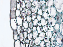
Cross section of collenchyma cells
Collenchyma is Greek word where "Collen" means gum and "chyma" means infusion. It is a living tissue of primary body like Parenchyma. Cells are thin-walled but possess thickening of cellulose, water and pectin substances (pectocellulose) at the corners where number of cells join together. This tissue gives a tensile strength to the plant and the cells are compactly arranged and have very little inter-cellular spaces. It occurs chiefly in hypodermis of stems and leaves. It is absent in monocots and in roots.Sometimes it contains chlorophyll which can help them photosynthesis.
Collenchymatous tissue acts as a supporting tissue in stems of young plants. It provides mechanical support, elasticity, and tensile strength to the plant body. It helps in manufacturing sugar and storing it as starch. It is present in the margin of leaves and resist tearing effect of the wind.

Cross section of collenchyma cells
Sclerenchyma
Sclerenchyma is Greek word where "Scleren" means hard and "chyma" means infusion. This tissue consists of thick-walled, dead cells and protoplasm is very less i.e. negligible. These cells have hard and extremely thick secondary walls due to uniform distribution and high secretion of lignin. Lignin deposition is so thick that the cell walls become strong, rigid and impermeable to water which is also known as stone cell or sclereids. This type of tissues are mainly of two divisions: 1) Sclerenchyma fiber and 2) Sclereids. Schlerenchyma cells have a narrow lumen and are long and narrow and unicellular.
Epidermis
The entire surface of the plant consists of a single layer of cells called epidermis or surface tissue. The entire surface of the plant has this outer layer of epidermis. Hence it is also called surface tissue. Most of the epidermal cells are relatively flat. The outer and lateral walls of the cell are often thicker than the inner walls. The cells forms a continuous sheet without inter cellular spaces. It protects all parts of the plant.
Complex permanent tissue
The complex tissue consists of more than one type of cells which work together as a unit. Complex tissues help in the transportation of organic material, water and minerals up and down the plants. That is why it is also known as conducting and vascular tissue. The common types of complex permanent tissue are:
- Xylem or wood
- Phloem or bast.
Xylem and phloem together form vascular bundles.
Xylem
Xylem consists of:
- Tracheid
- Vessel members
- Xylem fibers
- Xylem parenchyma
Xylem serves as a chief conducting tissue of vascular plants. It is responsible for the conduction of water and mineral ions/salt.

Cross section of 2 year old Tilia Americana, highlighting xylem ray shape and orientation.
Xylem tissue is organized in a tube-like fashion along the main axes of stems and roots. It consists of a combination of parenchyma cells, fibers, vessels, tracheids, and ray cells. Longer tubes made up of individual cells are vessels (tracheae), while vessel members are open at each end. Internally, there may be bars of wall material extending across the open space. These cells are joined end to end to form long tubes. Vessel members and tracheids are dead at maturity. Tracheids have thick secondary cell walls and are tapered at the ends. They do not have end openings such as the vessels. The tracheids ends overlap with each other, with pairs of pits present. The pit pairs allow water to pass from cell to cell.
Though most conduction in xylem tissue is vertical, lateral conduction along the diameter of a stem is facilitated via rays. [1] Rays are horizontal rows of long-living parenchyma cells that arise out of the vascular cambium. In trees and other woody plants, rays radiate out from the center of stems and roots, and appear like spokes on a wheel in cross section. Rays, unlike vessel members and tracheids, are alive at functional maturity. [2]
.jpg)
Cross section of 2 year old Tilia Americana, highlighting xylem ray shape and orientation.
Comments
Post a Comment